
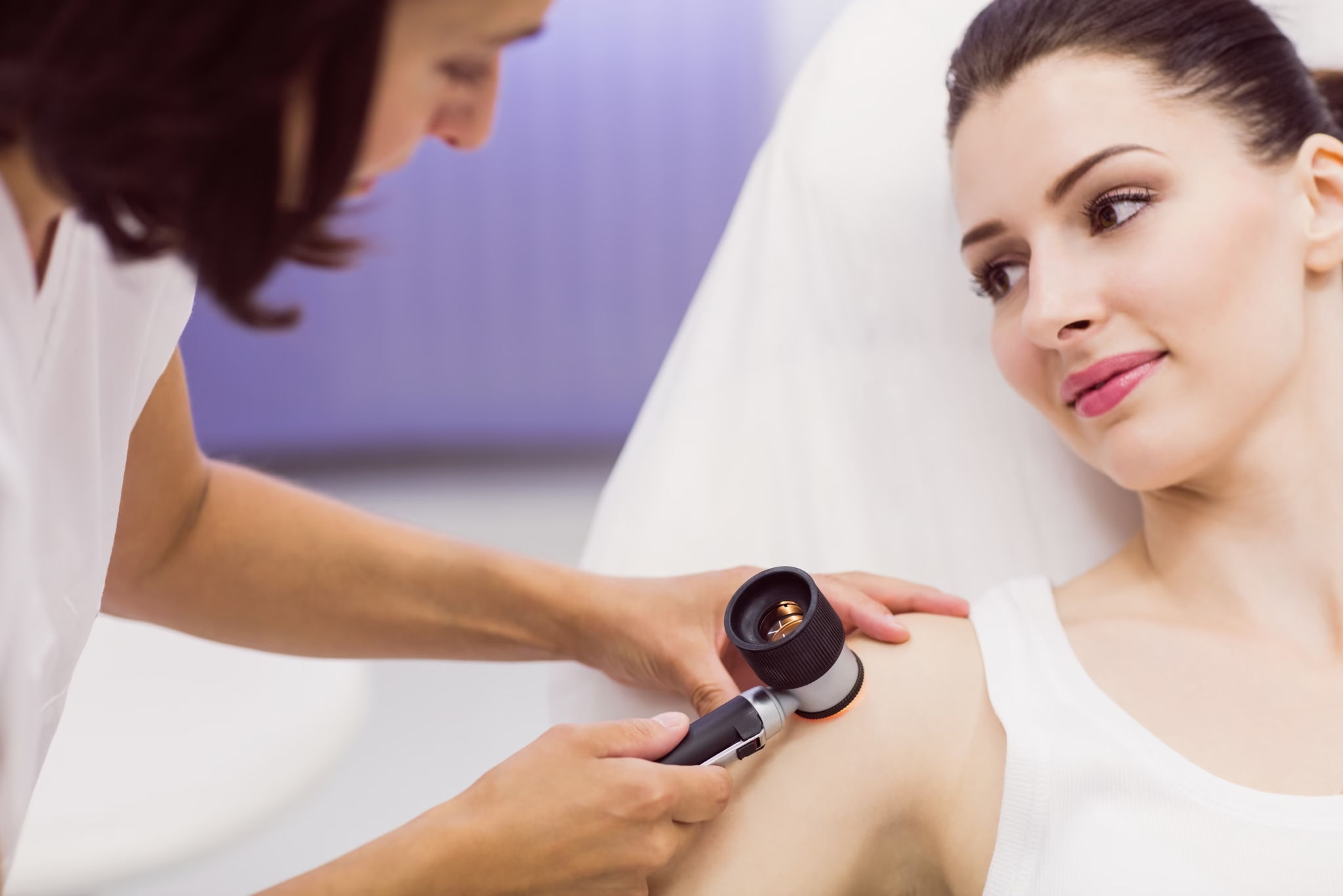
Dermoscopy is a non-invasive diagnostic method that allows a detailed examination of moles and other pigmented skin lesions using a special instrument – the dermoscope. The magnified image and special lighting allow the doctor to see structures beneath the surface of the skin that are not visible to the naked eye, significantly increasing the accuracy of the diagnosis.
This method is key to the early detection of melanoma and other skin tumours. Early detection of such changes significantly increases the success of treatment and gives the patient the best prognosis.
Who is this examination for?
A mole check with a dermoscope is recommended:
for people with a large number of moles
if there has been a change in the shape, colour, size or edges of existing moles
for people with a family history of melanoma
if you are often exposed to the sun or use tanning beds
if there are atypical changes or if moles itch, bleed or flake
What does the examination look like?
The examination is completely painless, short and requires no special preparation. The doctor visually examines all the moles and analyses in more detail those that are assessed as potentially risky. If needed, digital photographs can be taken to monitor changes over time.
If a malignant change is suspected, the patient immediately receives instructions for the next steps – further diagnostics, monitoring or removal of the lesion.
Our team of doctors combines experience, expertise and modern technology to provide you with the most reliable and safest diagnosis. We offer complete dermatological support – from the first examination and digital documentation to further treatment if needed.